Eye Defects (Congenital) in Dogs

Congenital Ocular Anomalies in Dogs
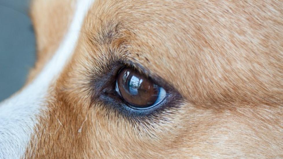
Congenital abnormalities of the eyeball or its surrounding tissue are generally evident shortly after a puppy's birth, but may develop within the first six to eights weeks of life. Most defects are genetically inherited; for example, persistent pupillary membrane (PPM), which occurs when strands of fetal tissue remain on the eye after birth, is more prone in Basenjis, Pembroke and Cardigan Welsh Corgis, chow chows, and mastiffs.
Meanwhile, persistent hyperplastic tunica vasculosa lentis (PHTVL) and persistent hyperplastic primary vitreous (PHPV) is most frequently inherited in Doberman pinschers. Multifocal retinal dysplasia (malformation of the retina) is found in English springer Spaniels; collie eye anomaly in collies, Shetland sheepdogs, and Australian shepherds; retinal dystrophy in Briards, photoreceptor dysplasia (malformation of the cells that perceive light and color) in Collies, Irish Setters, miniature schnauzers, and Norwegian elkhounds.
Ocular abnormalities can also develop spontaneously (e.g., colobomas of ther anterior) or occur in utero. Exposure to toxic compounds, lack of nutrients, and systemic infections and inflammations during pregnancy (such as panleukopenia) are other potential risk factors for ocular abnormalities.
Symptoms and Types
There are a variety of abnormalities that can affect a dog's eye or surrounding tissues. The following are some of the more common issues and their corresponding signs:
- Colobomas of the lid
- May appear as notch in eyelid, or tissue of the eyelid may be missing
- Variable eyelid twitching and watery eyes
- Colobomas of the iris
- Misshapen iris
- Sensitivity to bright light
- Does not typically affect vision
- Most common in herding dogs (i.e., Basenji, Collie, Australian sheepdog)
- Persistent pupillary membranes (PPM)
- Fetal tissue will remain on the eye after birth
- Variable iris defects
- Variable cataracts
- Variable colobomas of the uvea
- Common in Basenjis
- Dermoids
- Tumor-like cysts on eyelid(s) conjuctiva, or cornea
- Variable eyelid twitching and watery eyes
- Iris cysts
- Often not visible, as the cyst is located behind the iris
- May not have symptoms besides slight bulging of the iris, unless the cyst is interfering with the field of vision
- Congenital glaucoma (high pressure within the eye) with buphthalmos (abnormal enlargement of eyeball)
- Tearing
- Enlarged, red, and painful eye
- Congenital cataracts
- Cloudiness in the eyes
- Often inherited (e.g., Cavalier King Charles spaniels)
- Congenital keratoconjunctivitis sicca (KCS)
- Also referred as dry eye
- Common in Yorkshire terriers
- Other congenital issues
- Lack of pupils or abnormally-shaped pupil
- Lack of tear duct openings (Cocker Spaniels)
- Lack of iris
- Persistent hyperplastic tunica vasculosa lentis (PHTVL) and persistent hyperplastic primary vitreous (PHPV)
- Begins in utero, with progressive atrophy of the vascular system that supports the eye lens
- Common in Briards, Cocker Spaniels, beagles, rottweilers
- Retinal dysplasia
- Appears as folds or rosette shapes on the retina
- Common in Briards
- Retinal detachment
- Retina detaches from the back of the eye causing blindness
- Common in Labrador retrievers, Bedlingtons, and Sealyham terriers
- Photoreceptor dysplasia
- Night blindness (when rods are affected)
- Day blindness (when cones are affected)
- Slow or absent pupillary reflex to light (when pupil does not contract or dilate normally)
- Involuntary eye movement
- Optic nerve underdevelopment
- Often results in blindness
- Common in Miniature and toy poodles
- Rod-cone malformation
- Rod and cone malformation common in Irish setters and collies
- Rod malformation common in Norwegian elkhounds
- Cone malformation in Alaskan malamutes
In addition, hereditary defects, such as corneal opacities, PPM, cataracts, retinal detachement, and dysplasia, are often associated with the following factors:
- Abnormally small eyes
- Missing eyeball
- Hidden eyeball (due to other eye deformities)
Causes
- Genetic
- Spontaneous malformations
- Uterine conditions (e.g., infections and inflammations during pregnancy)
- Toxicity during pregnancy
- Nutritional deficiencies during pregnancy
Diagnosis
You will need to provide as much of your dog's medical history as you have available to you, such as in utero conditions (i.e., whether its mother was ill, her diet, etc.), and the dog's development and environment after birth. After taking a thorough history, your veterinarian will test the health of the eye.
A Schirmer tear test may be used to see if your dog's eyes are producing an adequate amount of tears. If high pressure in the eye (glaucoma) is suspected, a diagnostic tool called a tonometer will be applied to your dog's eye to measure its internal pressure. Abnormalities within the eye, meanwhile, will be examined with an indirect ophthalmoscope and/or a slitlamp biomicroscope.
An ultrasound of the eyes may also reveal problems with the lens of the eyeball, the vitreous humor (the clear fluid which fills the space between the lens and retina), the retina, or other problems that are taking place in the posterior (back) segment of the eye. In the case of iris cysts, ultrasound will help your doctor determine if the mass behind the iris is in fact a cyst or a tumor. Cysts do not always behave uniformly: some grow, while others shrink. In most cases follow-ups to check the progress of the cyst will be the extent of treatment, until further intervention is warranted.
Another useful diagnostic method called angiography can also be used for viewing problems in the posterior of the eye, such as detachment of the retina and abnormal blood vessels in the eye. In this method, a substance that is visible on X-ray (radiopaque) is injected into the area that needs to be visualized, so that the full course of blood vessels can be examined for irregularities.
Treatment
Treatment will depend on the specific type of eye abnormality that is affecting your dog. Depending on your veterinarian's experience with eye diseases, you may need further treatment with a trained veterinary ophthalmologist. Surgery can repair some congenital birth defects, and medicines can be used to mitigate the effects of some types of defects. Congenital keratoconjunctivitis sicca (KCS), commonly known as dry eye, can often be medically treated with tear substitutes in combination with antibiotics. Other medicines called mydriatics may be used to increase vision when congenital cataracts are present in the center of your dog's eye lenses.
In cases of photoreceptor dysplasia, there is no medical treatment that will delay or prevent its progress, but dogs with this condition generally do not suffer from any other physical abnormality and can learn to manage their environment very well, as long as they are able to depend on their environment being stable and safe.
Living and Management
Congenital KCS requires frequent checkups with a veterinarian to monitor tear production and the status of the external eye structures. Abnormalities such as congenital cataracts, PHTVL, and PHPV require checkups twice yearly to monitor progression.
In addition, since most congenital ocular anomalies are hereditary, you should not breed a dog that has been diagnosed with any of these disorders.