The Microscopic World of Veterinary Medicine


I think when many people think of a veterinarian, they think of observations and procedures performed at the macroscopic level: listening to a heart beat, suturing a leg wound, administering a vaccine. Indeed, most of the things I do as a large animal vet are done on the large scale. However, some very important diagnostics require the use of the trusty microscope, a wonderful tool which I think sometimes gets pushed into a dusty corner of the lab bench and scowled at by some.
I will allow that on the whole, large animal vets don’t seem to use the microscope as often as small animal vets, mostly due to differences in primary complaints of respective patients. One of the chief problems seen in a small animal clinic is ear infections that diagnostically will include a good ol’ swab and look-see under the microscope for bacteria and yeast. Small animal vets also diagnose a plethora of common skin problems under the microscope. Large animal vets rarely if ever do any type of ear swab (I don’t believe I ever have) since horses, cattle, sheep, and goats just don’t get yeasty ears like Labradors do, and our patients’ skin diseases don’t require “skin scrapes” quite as frequently. There is also the issue of convenience. Large animal vets on the road rarely have the luxury of a truck-side microscope and samples often have to wait in the truck until we get back to the office.
However, one of the most common uses of the microscope in both the small and large animal realms is the fecal float. A diagnostic tool that takes a small fecal sample, mixes it with a special solution, and then sits the resultant “poo-slurry” in a tube with a microscope coverslip on top, the fecal flotation test determines the presence of gastrointestinal parasites eggs.
There are many methods for the fecal flotation test. Variations include how much fecal matter you need, what type of solution you use (choices include sugar water and zinc sulfate, which cause the fecal eggs to separate from the fecal matter and rise to the top of the tube), whether or not you centrifuge the sample, and how long you let the sample sit before examining it under the microscope. Each variation has its pros and cons, including time and expertise required, expense, and sensitivity.
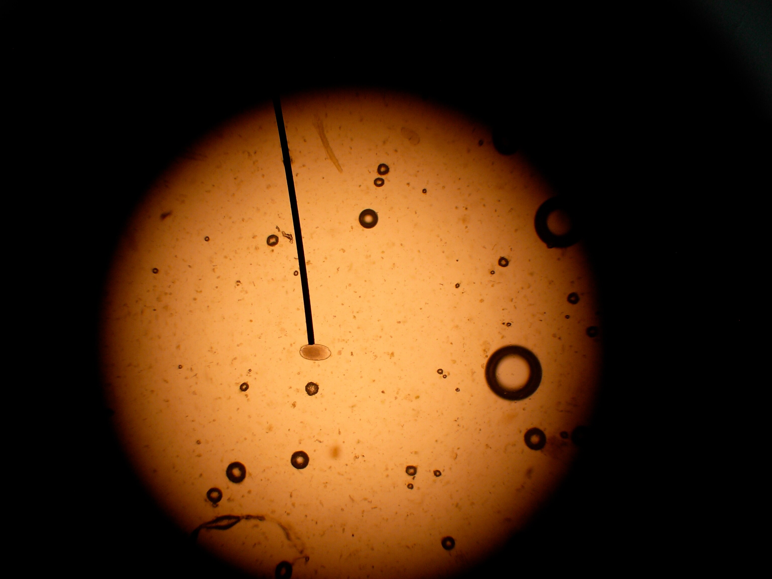
Once you’ve got the coverslip ready and on the slide, it’s then time to venture into the microscopic world of parasite eggs. Some find this boring. I’ll admit if I have twenty slides to read at one sitting, counting egg after egg after egg does get a bit tedious. But other times, I get lost in the world in front of my eyes. Air bubbles appear as large lenses, plant seeds can look like something out of a sci-fi novel, and occasionally you’ll get a misplaced grass mite or other bug in there, appearing amusingly enormous.
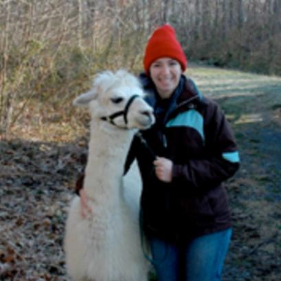
The identification of different types of parasite eggs takes time and training. Not only can different parasites have different looking eggs (although not always the case, as in a group of roundworms in horses, cattle, and small ruminants called strongyles), but also different species of animals carry different types of parasites. A harmful type of coccidia in alpacas looks slightly different than mostly harmless coccidia in sheep. However, with practice, egg identification becomes old hat and every so often, if I haven’t seen, say, a Nematordius egg in a while and I find one, it’s like visiting an old friend. Well, maybe not quite that nice, but I still get a friendly feeling of familiarity.

Dr. Anna O’Brien
Image: Nito / Shutterstock